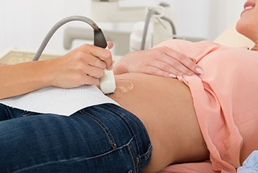
Ultrasound (also called sonography or ultrasonography) is a noninvasive imaging test. An ultrasound picture is called a sonogram. Ultrasound uses high-frequency sound waves to create real-time pictures or video of internal organs or other soft tissues, such as blood vessels.
Ultrasound enables healthcare providers to “see” details of soft tissues inside your body without making any incisions (cuts). And unlike X-rays, ultrasound doesn’t use radiation.Although most people associate ultrasound with pregnancy, healthcare providers use ultrasound for many different situations and to look at several different parts of the inside of your body.
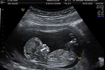
Book an AppointmentPregnancy dating scans, typically done at 6-12 weeks, use ultrasound to determine the due date. They help identify early fetal growth, number of pregnancies, and any potential complications.
The NT scan (nuchal translucency) measures fluid at the back of the fetal neck to assess the risk of chromosomal abnormalities like Down syndrome.
Growth scans monitor fetal development, assessing size, weight, and growth patterns to identify potential complications like intrauterine growth restriction.
Obstetric Dopplers use ultrasound to measure blood flow in the placenta and umbilical cord, assessing fetal health and detecting potential complications.
Biophysical profiles combine ultrasound and non-stress tests to assess fetal health, evaluating movement, muscle tone, breathing, and amniotic fluid levels.
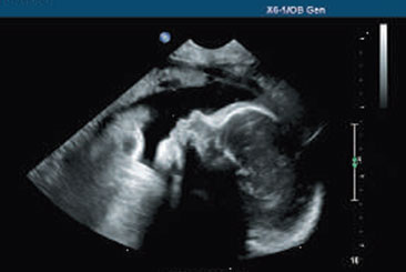
Pelvic ultrasound uses sound waves to visualize reproductive organs, diagnosing conditions like ovarian cysts, fibroids, or ectopic pregnancy.
A follicular study tracks ovarian follicle growth and ovulation, aiding in fertility treatments by monitoring egg maturation and timing.